Virtual Microscopy > Hematopoiesis and Lymphoid > Teaching Cases
Case 1: Normal Bone Marrow
Learning Objectives:
- Identify the morphologic features that characterize the various stages of erythroid development.
- Describe the role of bone marrow aspirate and biopsy to diagnose anemia.
- Describe the differentiation of committed erythroid progenitors from pluripotent stem cells.
Reading:
- Kierszenbaum AL: Chapter 6: Blood and Hematopoiesis. Histology and Cell Biology: An Introduction to Pathology. Mosby, Inc., St. Louis, 2002, pp. 147-150; 157-165.
- Aster JC: Chapter 13: Red Cells and Bleeding Disorders. In: Kumar V, Fausto N, Abbas AK: Robbins & Cotran Pathologic Basis of Disease. 7th ed., WB Saunders, Philadelphia, 2005, pp. 619-622.
Optional Reading:
- Riley RS, Hogan TF, Pavot DR, Forysthe R, Massey D, Smith E, Wright, Jr L, Ben-Ezra JM: A pathologist's perspective on bone marrow aspiration and biopsy: I: Performing a bone marrow examination. J Clin Lab Anal 2004; 18:70-90.
- Riley RS, Ben-Ezra JM, Pavot DR, et al: An Illustrated Guide to Performing the Bone Marrow Aspiration and Biopsy. Virginia Commonwealth University Department of Pathology (http://www.pathology.vcu.edu).
Clinical History:
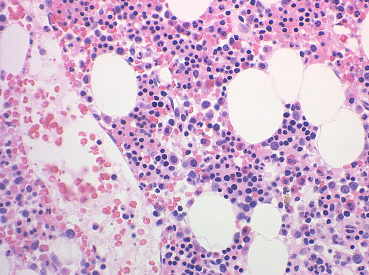
A bone marrow aspirate and biopsy were obtained from a 27 year old woman who volunteered to participate in a research study evaluating a new method for quantifying hematopoietic stem cells. The bone marrow cellularity and morphology were within normal limits. Images of the bone marrow aspirate smear and biopsy are available for review [Case1.1-Case1.11].
Protocol for Image Review:
- Examine the gross images of the marrow aspirate [Case1.01] and gross and low power images of the biopsy [Case1.02; Case1.03]. Identify bony spicules and the location of hematopoietic cells in each specimen.
- Using the microscopic images of the marrow aspirate [Case1.04, Case1.05, Case1.06, Case1.07], identify the following stages of erythroid maturation: proerythroblast, basophilic erythroblast; polychromatophilic erythroblast; orthochromatophilic erythroblast, erythrocyte.
- Using the microscopic images of the marrow biopsy [Case1.08, Case1.09, Case1.10, Case1.11], identify marrow adipose cells, bony trabeculae, fibroblasts, sinusoids, vascular endothelial cells, hemosiderin pigment, erythroid precursors, erythrocytes, myeloid precursors, and megakaryocytes.
Questions:
- Distinguish between the terms "red" and "yellow" bone marrow. Where is red bone marrow found in an adult?
- Describe the differences between a bone marrow aspirate and a bone marrow biopsy. What body sites are typically used for obtaining a bone marrow specimen for histologic examination in an adult? a child?
- Which stages of erythropoiesis retain the capacity for cell division?
- What is erythropoietin? At what stages of erythropoiesis does erythropoietin act?
- How can stem cells be identified in bone marrow specimens? What is a CFU-E?
Summary of photomicrographs for case 1:
- Bone marrow aspirate (Wright-Giemsa stain, gross)
- Bone marrow biopsy (Hematoxylin-Eosin stain, gross)
- Bone marrow biopsy (Hematoxylin-Eosin stain, 10x)
- Bone marrow aspirate (Wright-Giemsa stain, 100x)
- Bone marrow aspirate (Wright-Giemsa stain, 100x)
- Bone marrow aspirate (Wright-Giemsa stain, 100x)
- Bone marrow aspirate (Wright-Giemsa stain, 100x)
- Bone marrow biopsy (Hematoxylin-Eosin stain, 20x)
- Bone marrow biopsy (Hematoxylin-Eosin stain, 40x)
- Bone marrow biopsy (Hematoxylin-Eosin stain, 40x)
- Bone marrow biopsy (Hematoxylin-Eosin stain, 40x)
(back to top)
Case 2: Normal Peripheral Blood
Learning Objectives:
- Describe the morphologic features of normal erythrocytes (size, shape, volume, color).
- Define the following parameters in a complete blood count (CBC): hemoglobin, hematocrit, RBC count, MCV, MCH, MCHC, RDW. What are the reference ranges for these parameters?
- Define anemia.
Reading:
- Kierszenbaum AL: Chapter 6: Blood and Hematopoiesis. Histology and Cell Biology: An Introduction to Pathology. Mosby, Inc., St. Louis, 2002, pp. 147-150; 157-165.
- Aster JC: Chapter 13: Red Cells and Bleeding Disorders. In: Kumar V, Fausto N, Abbas AK: Robbins & Cotran Pathologic Basis of Disease. 7th ed., WB Saunders, Philadelphia, 2005, pp. 622-624.
Clinical History:
A 27 year old woman is referred to a family physician for a health screen prior to purchasing a $5,000,000 whole life insurance policy. The physical examination is unremarkable. A CBC is obtained as part of the evaluation, with the following results:
Protocol for Image Review:
- Case2.1 is a gross scan of a peripheral blood smear. The approximate field location for microscopic images (Case2.02, Case2.03, Case2.04) is marked by the end of each black line. Which microscopic field provides optimal RBC morphology? Why?
- Using images Case2.05 and Case2.06, identify the morphologic features of normal RBCs.
Questions:
- What are the expected normal ranges for the measured parameters in the CBC? Is this patient anemic?
- Describe the morphologic features of the erythrocytes. How much variation do you recognize in cell size, shape, and color?
- Are the erythrocytes normocytic, microcytic, or macrocytic? What is the normal lifespan of the circulating erythrocytes? What is a reticulocyte?
- What anticoagulant (tube color) is used to collect blood for a CBC? Why is an anticoagulant necessary? Describe the difference between serum and plasma.
Summary of photomicrographs for case 2:
- Peripheral blood (Wright-Giemsa stain, gross)
- Peripheral blood (Wright-Giemsa stain, 20x)
- Peripheral blood (Wright-Giemsa stain, 100x)
- Peripheral blood (Wright-Giemsa stain, 20x)
- Peripheral blood (Wright-Giemsa stain, 100x)
- Peripheral blood (Wright-Giemsa stain, 100x)
(back to top)
Case 3: Hereditary Elliptocytosis
Learning Objectives:
- Describe the normal components of the cell membrane of an erythrocyte.
- Recognize morphologic disease entities associated with RBC cytoskeletal defects.
Reading:
- Kierszenbaum AL: Chapter 6: Blood and Hematopoiesis. Histology and Cell Biology: An Introduction to Pathology. Mosby, Inc., St. Louis, 2002, pp. 147-150; 157-165.
Clinical History:
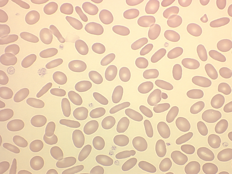
A peripheral blood smear was obtained from an asymptomatic 45 year old female medical technologist. The results of the CBC (erythroid parameters only) are listed below:
Protocol for Image Review:
- Review the RBC morphology in images (case3.01, case3.02, case3.03, case3.04) and contrast your findings with what you have seen in Case 2.
Questions:
- Describe the morphology of this patient's erythrocytes.
- What is the basis for the abnormal RBC morphology in this case?
- What is the clinical significance of this abnormality? Would this abnormality be detected in a routine CBC?
- What clinicopathologic entity results from a deficiency of spectrin in the RBC membrane?
Summary of photomicrographs for case 3:
- Peripheral blood (Wright-Giemsa stain, 100x)
- Peripheral blood (Wright-Giemsa stain, 100x)
- Peripheral blood (Wright-Giemsa stain, 100x)
- Peripheral blood (Wright-Giemsa stain, 100x)
(back to top)
Case 4: Hemoglobinopathy (Hb SS)
Learning Objectives:
- Describe erythrocyte abnormalities associated with sickle cell disease as an example of a disease caused by a defective hemoglobin molecule.
- Correlate abnormal cell morphology with predicted clinical features.
Reading:
- Kierszenbaum AL: Chapter 6: Blood and Hematopoiesis. Histology and Cell Biology: An Introduction to Pathology. Mosby, Inc., St. Louis, 2002, pp. 147-150; 157-165.
- Aster JC: Chapter 13: Red Cells and Bleeding Disorders. In: Kumar V, Fausto N, Abbas AK: Robbins & Cotran Pathologic Basis of Disease. 7th ed., WB Saunders, Philadelphia, 2005, pp. 628-632.
Clinical History:
A 60 year old African-American male was admitted for surgery for a leg ulcer. A CBC was requested as part of the pre-operative work-up. This showed (erythroid parameters only):
| RBC: | 2.18 x 10¹²/L |
| Hgb: | 7.0 g/dL |
| Hct: | 20.0 % |
| MCV: | 91.7 fL |
| MCH: | 32.1 pg |
| MCHC: | 35.0 g/dL |
| RDW: | 20.8 % |
Images of the peripheral blood smear are available for review [Case4.01, Case4.02, Case4.03, Case4.04, Case4.05].
Protocol for Image Review:
- Identify the following: sickled RBC, Howell-Jolly body, polychromatophilic RBC, platelet, target cell.
Questions:
- Describe the abnormal RBC morphology in this case.
- What is the molecular defect in this case?
- What clinical consequences might arise as a result of the abnormal RBC morphology illustrated here?
- What considerations might be necessary before the patient can be cleared for surgery?
Summary of photomicrographs for case 4:
- Peripheral blood (Wright-Giemsa stain, 100x)
- Peripheral blood (Wright-Giemsa stain, 100x)
- Peripheral blood (Wright-Giemsa stain, 100x)
- Peripheral blood (Wright-Giemsa stain, 100x)
- Peripheral blood (Wright-Giemsa stain, 100x)
(back to top)
Case 5: Megaloblastic anemia
Learning Objectives:
- Describe the appearance of megaloblastic erythroid maturation.
- Understand the basis for the nuclear:cytoplasmic dyssynchrony that is characteristic of this entity.
Reading:
- Kierszenbaum AL: Chapter 6: Blood and Hematopoiesis. Histology and Cell Biology: An Introduction to Pathology. Mosby, Inc., St. Louis, 2002, pp. 147-150; 157-165.
- Aster JC: Chapter 13: Red Cells and Bleeding Disorders. In: Kumar V, Fausto N, Abbas AK: Robbins & Cotran Pathologic Basis of Disease. 7th ed., WB Saunders, Philadelphia, 2005, pp. 638-643.
Clinical History:
A 55 year old woman with history of chronic alcoholism was admitted for severe anemia and fatigue. Physical examination showed mild jaundice, an enlarged, tender liver and an enlarged spleen. Laboratory studies showed elevated GGT, alkaline phosphatase, and bilirubin. A CBC showed the following results (erythroid parameters only):
| RBC: | 1.16 x 10¹²/L |
| Hgb: | 5.0 g/dL |
| Hct: | 15.0 % |
| MCV: | 129 fL |
| MCH: | 43.1 pg |
| MCHC: | 33.3 g/dL |
| RDW: | 18.5 % |
Additional laboratory testing and bone marrow examination were performed to establish a diagnosis. Images of the bone marrow aspirate and biopsy are available for review [
Case5.01, Case5.02, Case5.03, Case5.04, Case5.05, Case5.06, Case5.07].
Protocol for Image Review:
- Using the images of the bone marrow aspirate (Case5.1-5.6), identify the following: megaloblastic maturation in erythroid precursors (proerythroblast, basophilic erythroblast, polychromatophilic erythroblast, orthochromatophilic erythroblast), nuclear:cytoplasmic dyssynchrony, mitotic figure. Compare the morphology with Case 1.
Questions:
- List morphologic features in peripheral blood and bone marrow erythroid precursors associated with megaloblastic maturation. Compare with Cases 1 & 2.
- Describe the defect that is associated with megaloblastic anemia. Why does this result in anemia?
- Distinguish the terms "megaloblastic anemia" from "macrocytic anemia." Are all macrocytic anemias megaloblastic?
- The cause of the patient's megaloblastic anemia in this case was folate deficiency, as confirmed by assay for serum folate: 1.6 ng/mL (N: 3-22 ng/mL) and RBC folate: 100 ng/mL (N: 174-665 ng/mL). Serum B12 was 787 pg/mL (N: 218-959 pg/mL). Given the etiology of the anemia in this case, what other cytologic changes might be noted in other tissues?
Summary of photomicrographs for case 5:
- Bone marrow aspirate (Wright-Giemsa stain, 100x)
- Bone marrow aspirate (Wright-Giemsa stain, 100x)
- Bone marrow aspirate (Wright-Giemsa stain, 100x)
- Bone marrow aspirate (Wright-Giemsa stain, 100x)
- Bone marrow aspirate (Wright-Giemsa stain, 100x)
- Bone marrow aspirate (Wright-Giemsa stain, 100x)
- Bone marrow biopsy (Hematoxylin-Eosin stain, 40x)
(back to top)


